- Created by PostDICOM.jpg)
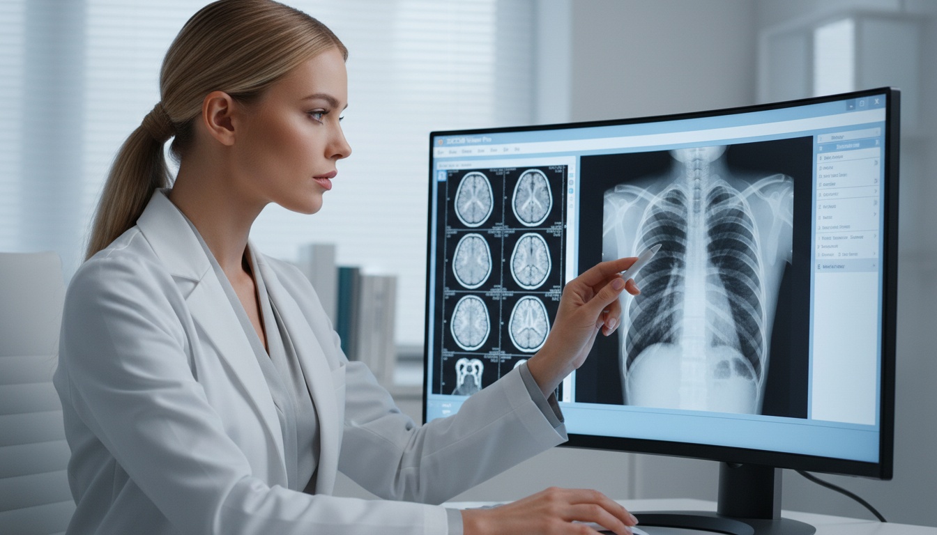
Medical imaging has been a further developmental step to seeing fixed CT, MRI, and X-ray images. Modern radiologists, experts, and healthcare teams require interactive solutions that could assist them to study anatomy in various directions, isolate structures, and cooperate remotely without being bound to specific workstations.
It is in this case that advanced image processing is needed.
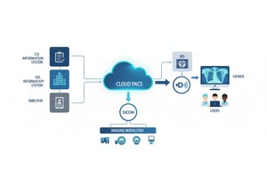
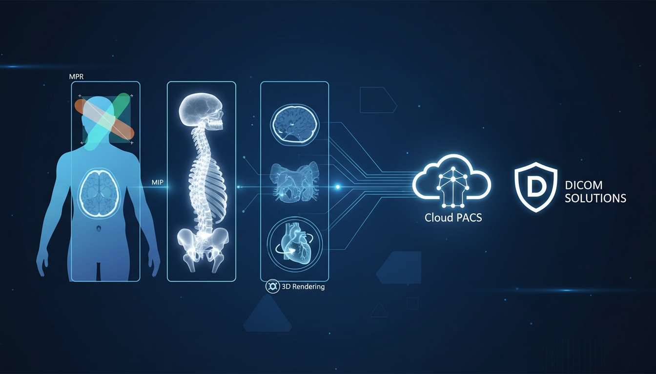
PostDICOM integrates cloud-based access to images with robust visualization, like Multiplanar Reconstruction (MPR), Maximum Intensity Projection (MIP), and 3D Rendering. The capabilities can assist clinicians to interpret studies more quickly, enhance diagnostic confidence, and facilitate modern remote imaging processes.
The sophisticated visualization tools (MPR, MIP and 3D rendering) are applied in the analysis of DICOM studies (CT and MRI). MPR recreates images in several planes in the anatomy, MIP in high-density structures such as vessels or bone, and 3D rendering produces volumetric representations to support surgical planning and teaching. PostDICOM provides these tools as a secure web browser without intricate local installation.
Conventional slice-by-slice imaging may be laborious, particularly in assessing complicated anatomy or fine pathology. Premier reconstruction tools assist clinicians to get out of the two-dimensional axial imaging and examine information more effectively.
This is important since healthcare organizations are becoming more in need of:
• Reduced Reporting Turnaround.
• Improved Inter-locational Cooperation.
• Improved Pre-surgical Planning
• More Precise Lesion And Vessel Analysis.
• Home, Clinic, And Partner Hospital Access.
• Less Reliance On Costly Local Workstations.
Cloud-based imaging systems are assisting in bridging that divide.
Multiplanar Reconstruction transforms a volumetric dataset into a number of viewing planes. Rather than being confined to the scan direction, the clinicians can view images in:
• Axial Plane
• Coronal Plane
• Sagittal Plane
• Oblique Custom Angles
Several anatomical discoveries are more distinguishable in a different plane than the acquisition position. As an example, spinal alignment, sinu anatomy, fracture, abdominal structures tend to be better revealed either in the coronary or sagittal view.
• Review Of Spine Disc Herniation.
• Sinus Ct Evaluation
• Orthopedic Trauma Imaging
• Abdominal Lesion Localization
• Review Of Cardiac And Vascular Path.
• Mri Soft Tissue Examination.
PostDICOM allows users to interactively manipulate planes on the web, which allows radiologists and other experts to view anatomy without exported reformats.
Maximum Intensity Projection shows the brightest voxels of a chosen slab of the volume. It is particularly helpful in analyzing high signal or dense structures.
MIP has applications in:
• Ct Angiography
• Pulmonary Vessel Review
• Mri Angiography
• Workflows Of Lung Nodules Detection.
• Emphasis On Bone Structure In The Chosen Studies.
Clinicians can easily highlight vessels or thick structures in a condensed view, rather than scrolling through hundreds of slices. This is able to accelerate the triage process and enhance the visibility of certain anatomy.
MIP can help a physician who is reviewing vascular stenosis to more clearly see vessel continuity and vessel narrowing patterns.
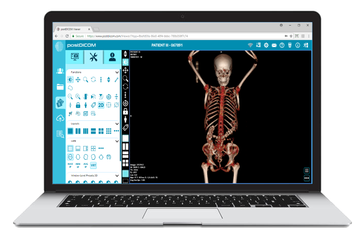
The 3D rendering converts the image datasets into volumetric models that recreate the anatomical depth and shape. Users are able to rotate and examine anatomy in 3D, rather than just looking at slices
Three-dimensional views are often valuable for:
• Surgical Planning
• Orthopedic Fracture Mapping
• Craniofacial Reconstruction Review
• Tumor Relationship Assessment
• Patient Education
• Referring Physician Communication
The complex structures may be more readily comprehended in 3D than in serial 2D slices. This assists not only the specialists but non-radiologist clinicians to interpret findings more confidently.
| Tool | Primary Purpose | Best For | Key Benefit |
| MPR | Multi-plane reconstruction | CT/MRI anatomy review | Better orientation |
| MIP | Highlight brightest voxels | Vessels, lungs, dense structures | Faster detection |
| 3D Rendering | Volumetric anatomy model | Surgery, ortho, communication | Realistic visualization |
 - Presented by PostDICOM.jpg)
In many cases, clinicians use all three tools during the same case review.
In the past, sophisticated image processing involved costly workstations locally and special software. The distributed healthcare teams are put under strain because of that model.
Modern healthcare systems need:
• Remote Radiology Access
• Teleradiology ReadingenvironmentsTeleradiology reading
• Multi-site Collaboration
• Faster Specialist Consultation
• Lower It Overhead
• Easier Deployment And Updates
PostDICOM addresses these needs by bringing advanced visualization into a web-based environment.
| Feature | Cloud-Based Viewer | Traditional Workstation |
| Access Location | Browser from approved locations | Fixed device or office network |
| Software Updates | Centralized | Manual / local installs |
| Collaboration | Easier remote sharing | Often slower or fragmented |
| Scalability | Flexible growth | Hardware dependent |
| IT Maintenance | Lower complexity | Higher support burden |
The move towards browser-based imaging is not just a matter of convenience to many healthcare organizations. It is also concerned with efficiency in operations, accelerated access and ease of management of infrastructure.
With a compatible browser, users are able to access studies and advanced tools.
The same case can be reviewed more effectively by clinicians in various locations.
Cloud systems are able to minimize the reliance on workstation-based deployments.
Studies can be accessed by radiologists, surgeons and experts either at office, clinic or even at distant locations, depending on policy controls.
With a larger imaging volume, organizations are able to grow more efficiently than the previous workstation-only designs.
Consider an ED that examines a patient with a complex case of face injuries due to an accident. The radiology team can initially scan the injury using standard CT slices, and proceed to scan fractures through MPR views in coronal and sagittal views. MIP can be used to emphasize thick bony structures to be visualized more quickly, whereas 3D rendering may give a better model of the fracture displacement to be used by the surgeon planning the intervention.
Rather than image export to various systems, the availability of these tools into a single viewer of a secure browser can enhance the communication between the radiologists, trauma physicians and surgical teams.
State-of-the-art image processing is useful in various fields of medicine. Although radiologists are the most likely to be the users, the advantages are also clearer visualization of the anatomy and quicker access to reconstructed images by surgeons, specialists, and referring clinicians.
| Specialty | How MPR, MIP, and 3D Rendering Help |
| Radiology | Improve CT and MRI interpretation, lesion localization, and cross-sectional review. |
| Orthopedics | Support fracture mapping, implant planning, alignment review, and surgical preparation. |
| Neurology / Neurosurgery | Assist with vascular studies, brain anatomy review, and structural planning. |
| Cardiology | Improve cardiac CT visualization and vessel pathway assessment. |
| Oncology | Help evaluate tumor extent and anatomical relationships. |
| ENT | Useful for sinus CT review, facial structures, and surgical planning. |
| Emergency Medicine | Faster review of trauma imaging and complex injury anatomy. |
This makes access to high-end browser-based visualization tools more valuable as additional specialties are dependent on imaging-guided decisions.
Remote reading environments have become widely used in North America. Many organizations are no longer able to do without advanced tools.
Radiologists expect:
• Fast Image Loading
• Smooth Multiplanar Navigation
• Diagnostic-quality Viewing
• Secure Access Controls
• Collaboration Features
• Anywhere Accessibility
Storage, sharing, and enhanced viewing platforms can enhance efficiency in operations.
When comparing cloud-based medical imaging solutions, organizations must consider:
• Data Encryption
• Access Permissions
• Audit Logs
• Secure Sharing Workflows
• Backup Architecture
• Regulatory Alignment For Their Region
High-tech tools are important, but business security is equally important.
In comparing vendors, inquire:
1. Does It Natively Support Mpr, Mip And 3d Rendering?
2. Does It Have A Fast Enough Browser Performance To Support Clinical Workflow?
3. Is It Secure To Have Multiple Users?
4. Are Workstation Software Easier To Deploy?
5. Is It Interoperable With Pacs / Ris / Emr?
6. Is It Scalable To Multiple Facilities?
PostDICOM is an all-in-one cloud imaging accessibility and advanced viewer platform. Healthcare teams can more effectively centralize workflow, rather than dividing archive, viewer, and collaboration tools.
It can be particularly useful to:
• Growing Imaging Centers
• Multi-location Clinics
• Teleradiology Groups
• Hospitals Upgrading Old Systems.
• Experts Who Require Quick Access To External Studies
Yes. MPR is often applied to CT and MRI data that have volumetric image data.
No. Although angiography is a typical application, depending on the study, MIP can be utilized to visualize lungs, bone, and other dense structures.
It is capable of aiding diagnosis, planning and communication. It is still subject to interpretation based on complete clinical analysis and source images.
In numerous scenarios, browser-based views can eliminate reliance upon local workstations. Precise appropriateness is based on clinical needs and organization guidelines.
They enhance accessibility, collaboration, scalability, and operational efficiency.
Yes. Advanced visualization is usually beneficial to surgeons, orthopedists, cardiologists, oncologists and referring physicians.
Hospitals, imaging groups, and specialties tend to have more than one location. In the case of teams that use different local systems, teamwork and access to images can be sluggish and intermittent. The cloud based platform having sophisticated viewing tools assists in standardizing workflow, minimize delays and provide clinicians with a more integrated experience across locations.
The ability to access scalably MPR, MIP and 3D visualization can become a strategic benefit and not merely a technical option as the demand of imaging continues to increase.
More sophisticated image processing is no longer just a luxury of reading rooms. The current healthcare teams should have intelligent and easily accessible tools that will help in making faster decisions and collaborating better.
Providing MPR, MIP, and 3D rendering over a secure web-based platform, PostDICOM allows organizations to modernize imaging processes and enhance usability in clinicians in multiple locations.
Advanced visualization is not something that can be included in the decision later, it is something that must be incorporated in the decision, should your team be considering cloud imaging platforms.
|
Cloud PACS and Online DICOM ViewerUpload DICOM images and clinical documents to PostDICOM servers. Store, view, collaborate, and share your medical imaging files. |